Research
Philosophy
People first. This is the guiding philosophy for our laboratory. Our greatest assets are the people in our lab, our collaborators, and the innovative ideas and enthusiasm they bring to our science. And, of course, the ultimate goal of our efforts is to help people; our patients with severe neurological disease and the loved ones caring for them.
Scientific Mission
We have developed human stem cell derived brain organoid models that are capable of generating complex physiological activities together with tools to record and quantify these. The core scientific mission of this laboratory is to continue to innovate and expand on what we have already developed while leveraging our current methodologies to dive deeper into the mechanisms of normal neural circuit development and how neurological disease perturbs circuit formation and function.
Overview
In all projects described below we primarily use organoids generated from iPSCs harboring single gene variants associated with epilepsy and autism. More recently, we have also acquired multiple iPSC lines harboring pathogenic mutations in the microtuble-associated protein tau (MAPT) gene. Patients with this mutation have an early onset Alzheimer’s-like dementia, and we are actively generating organoids harboring this mutation in our studies of hippocampal circuit formation and function.
Project Examples
The following represent a few (but not all) of the ongoing projects in the lab. We also spend considerable effort within all of our projects in the refinement and development of our organoid generation techniques and analysis approaches.
Using brain-region specific organoids to delineate regional divergence in human neural circuit development and function
Many neurological disorders are associated with impairments in multiple cognitive domains. For example, a patient with a primary diagnosis of autism may also have comorbid epilepsy and intellectual disability. Even in cases where the neurological disorder can be traced to a single gene pathogenic variant it remains unclear if a single upstream pathological driver has shared or unique effects on circuit function in distinct brain regions. This kind of regional divergence may have important implications for both disease pathogenesis and treatment. In recent work, we have focused on the differential impact of single gene variants on the development and function of hippocampal versus forebrain cortical circuits.
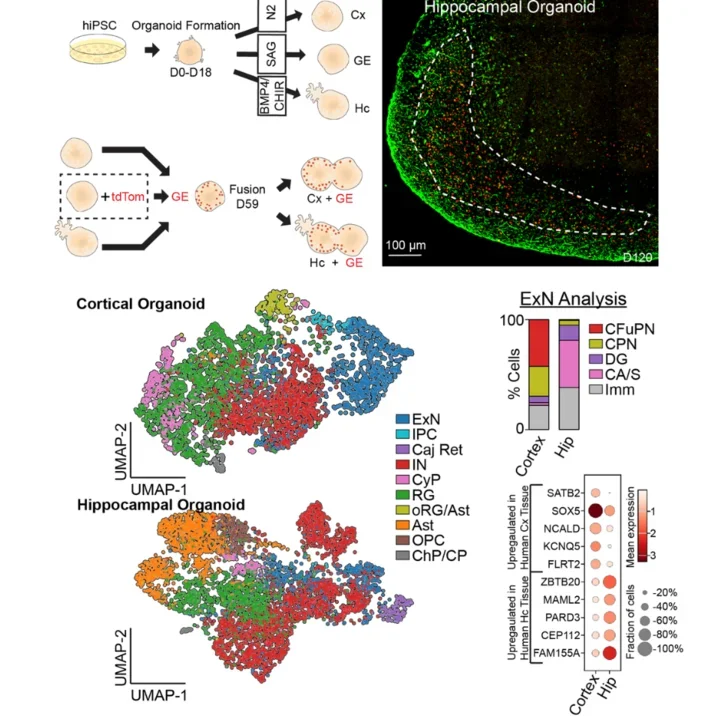
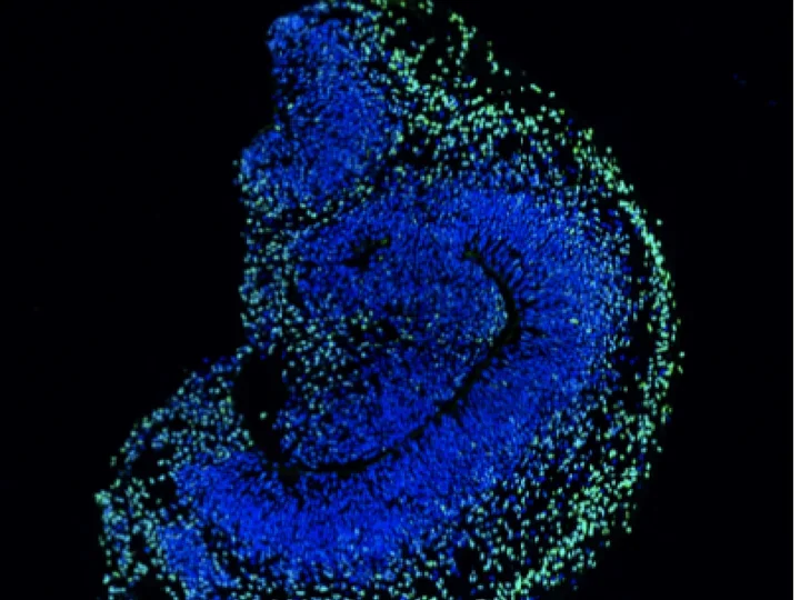
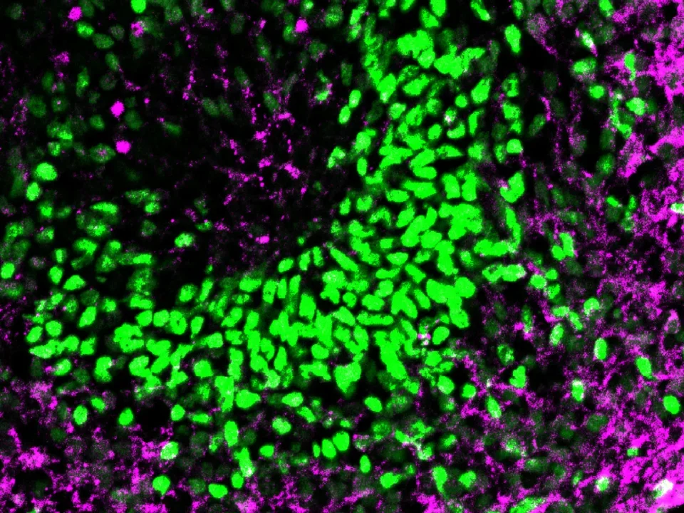
Figure Description: Top left, a schematic illustrating how we differentiate hippocampal versus cortical organoids and assembloids. Top right, an immunofluorescence image of a 120-day old hippocampal organoid showing canonical hippocampal cell types: Prox1+ dentate granule cells (in red) and KAI+ CA3 cells (in green). Bottom, multiple panels showing UMAP representations of cortical and hippocampal assembloid single cell RNA sequencing data. The bottom right has a bar graph demonstrating DG/CA-S like excitatory neurons in hippocampus versus CFuPN and CPN like excitatory neurons in cortex. Directly below this is a dot plot showing overlap of key markers from human hippocampal tissue with our hippocampal assembloid sequencing data, and similar overlap for human ex vivo cortex and our cortical assembloids.
Understanding network electrodynamics in developing brain microcircuits
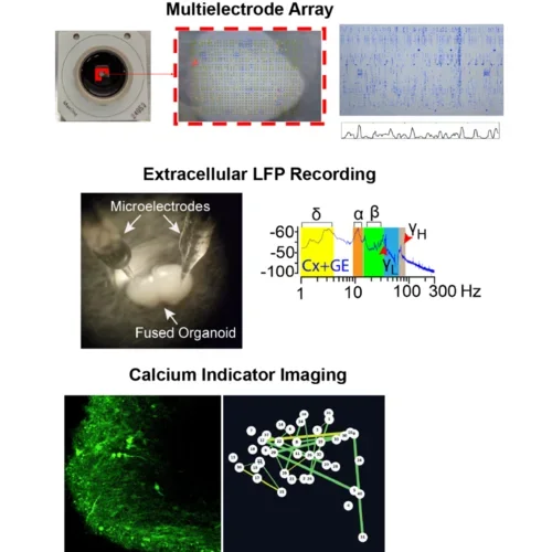
We use multiple, complementary, electrophysiologic approaches in brain organoids and assembloids to understand network electrophysiology. Multielectrode arrays provide impressive temporal data over wide areas, extracellular recordings provide data on neural oscillations that can be correlated with in vivo human data like EEGs, and calcium indicator imaging provide precise spatial information.
Figure Description: Images showing examples of the three main electrophysiology recording approaches used by our lab. Top, an image of an organoid on an MEA chip and its output. Middle, an image of an organoid subjected to microelectrode-based LFP recording and the resulting oscillatory data. Bottom, an example of an organoid subjected to calcium indicator imaging and a network connectivity map resulting from these data.
An Organoid Model of Hippocampal Circuit Function
We have generated hippocampal assembloids that can generate canonical hippocampus-associated neural circuit activities. For example, we have observed stereotyped patterns of theta-gamma phase amplitude coupling in control assembloids that are altered in variants harboring epilepsy and autism associated mutations. In ongoing projects, we have directly compared these oscillations to those taken directly from human patient recordings and found overlap.
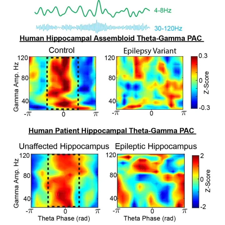
Figure Description: Top, Images of theta and gamma filtered oscillations taken from a hippocampal assembloid. Middle, representative morlet plots showing theta-gamma phase amplitude coupling in control and mutant hippocampal assembloids. The mutant harbors a variant associated with severe epilepsy. Bottom, the identical analysis applied to a human hippocampal intracranial recording showing similar patters of theta-gamma coupling in normal versus epileptic hippocampus. (Human recordings and data courtesy of Dr. Jack Lin, UC Davis)